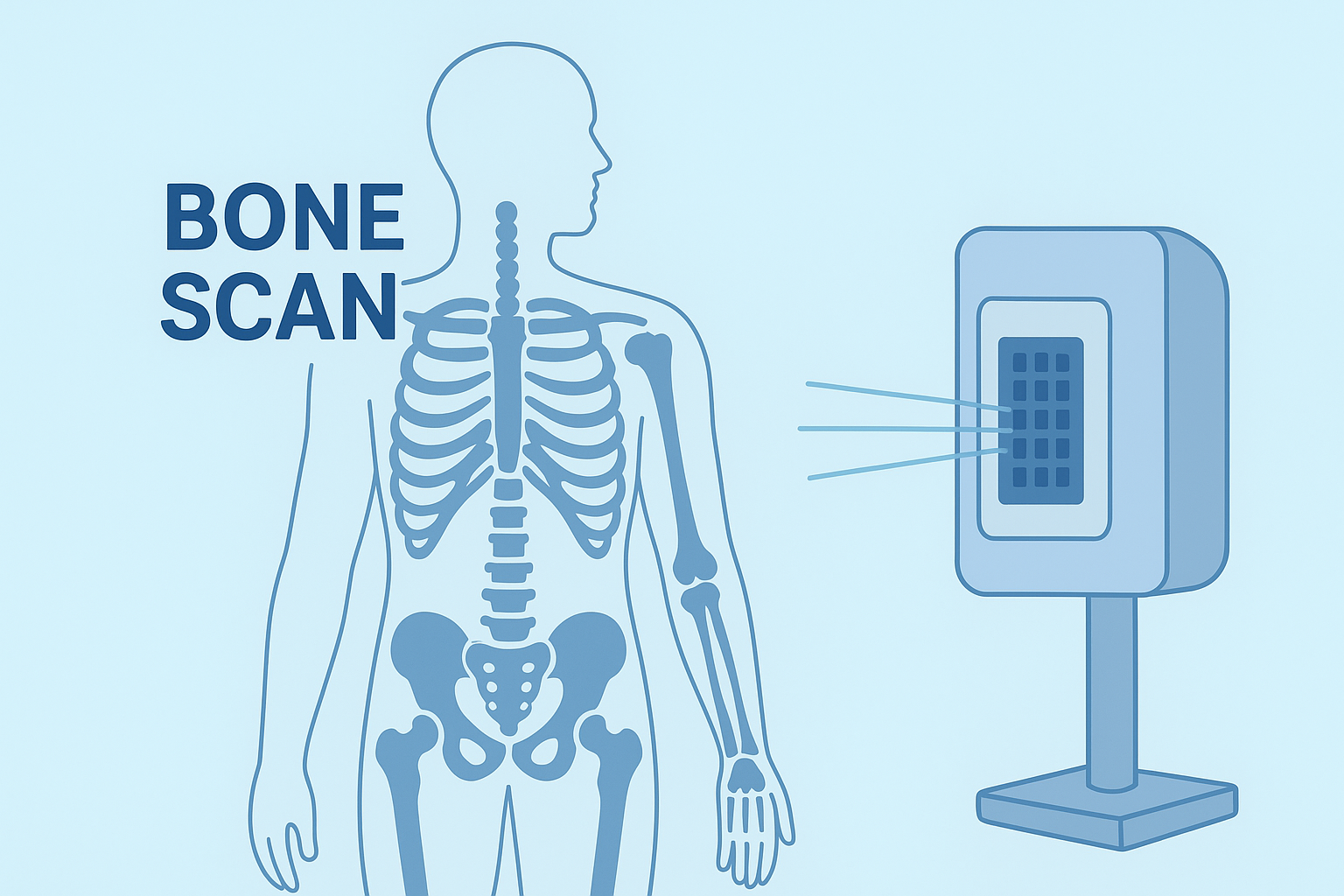
Bone Scan – Accurate Detection of Bone Disorders and Injuries
A Bone Scan is a highly sensitive nuclear medicine imaging test used to detect bone abnormalities that may not appear on traditional X-rays. As one of the leading centers for Bone Scan Nuclear Medicine Delhi, DIFI Healthcare helps in the early diagnosis of fractures, infections, arthritis, and bone tumors, allowing doctors to plan effective treatments before conditions worsen. We use advanced gamma camera technology and Technetium-99m tracers to provide detailed and accurate images of bone activity. Our expert nuclear medicine specialists ensure safe, precise, and comfortable scanning experiences for every patient.
Preparation Guidelines for a Bone Scan
To ensure the best imaging results, please follow these simple steps:
- Hydrate Well: Drink plenty of water before and after the scan to help flush out the tracer.
- Diet: No fasting is usually required — you can eat normally before the procedure.
- Medications: Inform your doctor about any ongoing medications.
- Pregnancy & Breastfeeding: Notify the medical team if you are pregnant or nursing.
- Comfortable Clothing: Wear loose, comfortable clothes without metal fasteners or jewelry.
Key Benefits of a Bone
Scan
- Early Detection: Identifies bone abnormalities at the earliest stage.
- Highly Sensitive: Detects small lesions or infections that X-rays may miss.
- Non-Invasive & Safe: Uses a small, safe amount of radioactive material.
- Guides Treatment Planning: Helps doctors evaluate bone healing, infection, or cancer spread.
- Quick & Comfortable: The entire procedure is painless and requires minimal preparation.
Bone scan nuclear medicine Delhi, Meerut & Dehradun
Purpose of a Bone Scan
A Bone Scan is performed to detect and monitor various bone-related conditions, including:
- Fractures (especially stress or hidden fractures not visible on X-rays)
- Bone Infections (Osteomyelitis)
- Arthritis and Joint Inflammation
- Bone Tumors or Metastasis (spread of cancer to bones)
- Unexplained Bone Pain or Swelling
- Post-Surgical Bone Evaluation
This test helps determine abnormal bone metabolism, highlighting areas of rapid bone growth or repair that may indicate disease or injury.

How a Bone Scan is Performed
- A small dose of Technetium-99m radiotracer is injected into your bloodstream.
- The tracer travels to your bones and highlights areas with unusual activity.
- After a short waiting period (2–4 hours), a gamma camera captures detailed images of your skeletal system.
- The entire process typically takes 3–4 hours, including the waiting and scanning time.
- Areas with increased tracer uptake indicate possible infection, fracture, or tumor activity, helping your doctor make an accurate diagnosis.
Why Choose DIFI Healthcare for Your Bone Scan?
- Advanced Gamma Camera Technology for superior imaging precision.
- Expert Nuclear Medicine Specialists with years of diagnostic experience.
- Safe, Hygienic & Patient-Centered Environment for maximum comfort.
- Fast, Reliable Reporting with accurate diagnosis and timely delivery.
- Affordable Scan Packages without compromising quality.
After the Scan
- You can resume normal activities immediately.
- Drink extra fluids to eliminate the radioactive tracer naturally.
- The tracer is completely safe and exits the body within 24 hours through urine.
Book Your Bone Scan at DIFI Healthcare
- Detect bone conditions early with a Bone Scan at DIFI Healthcare.
- Our expert imaging team ensures precise, safe, and comfortable diagnostic services to help you get the right treatment at the right time.
- Call us today or book your Bone Scan online at www.difi.in for accurate and affordable nuclear medicine imaging.
